Key (online version)
|
|
1a |
| One eye on each side of the head. |
|
4 |
| b |
| Both eyes on the same side of the head. |
|
2 |
| |
|
2a |
|
3 |
| b |
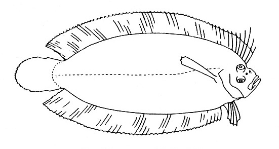
| Pectoral fins absent. [Fig. 394] |
| Soleidae |
|
|
| |
|
3a |
| Eyes on the right side of the fish's head. [Fig. 361] |
| Samaridae |
|
|
| b |
| Eyes on the left side of the fish's head. [Fig. 362] |
| Bothidae |
|
|
| |
|
4a |
| Either the gill openings are in front of the pectoral bases or the pectoral fins are absent. |
|
5 |
| b |
| Gill openings behind the level of the pectoral bases. Two short, blunt, stout dorsal spines on the head. [Fig. 330] |
| Antennariidae |
|
|
| |
|
5a |
| Pelvic fins present, though sometimes rudimentary or highly modified. |
|
6 |
| b |
| Pelvic fins totally absent. |
|
19 |
| |
|
6a |
| The 2 pelvic fins separate from one another. |
|
7 |
| b |
| The innermost rays of the 2 pelvic fins attached to one another for their full length by membrane. [Fig. 343] |
| Gobiidae (in part) |
|
|
| |
|
7a |
| Pelvic fins with 5 or fewer soft rays. |
|
8 |
| b |
| Pelvic fins with more than 5 soft rays. |
|
9 |
| |
|
8a |
| Dorsal fin composed of 2 or more completely separate parts. |
|
29 |
| b |
| A single dorsal fin, which may be more or less subdivided, but if so the sections are connected by at least a basal membrane. |
|
41 |
| |
|
9a |
| Anal fin with 4 spines. Dorsal fin with 10 or more spines, the base of the spinous portion longer than the base of the soft portion. [Fig. 341] |
| Holocentridae |
|
|
| b |
| No stiff, sharp spines at the front of the anal fin. |
|
10 |
| |
|
10a |
| Adipose fin present. Mouth extending well beyond the posterior border of the eye. [Fig. 331] |
| Synodontidae |
|
|
| b |
|
11 |
| |
|
11a |
| Snout elongate, tubular, with a small mouth at the tip. |
|
12 |
| b |
| Snout not in the form of an elongate tube with a small mouth at its tip. |
|
13 |
| |
|
12a |
| Soft dorsal preceded by a series of small, free spines; chin with a barbel; caudal without a median filament. Body compressed. [Fig. 332] |
| Aulostomidae |
|
|
| b |
| No free spines along back; no barbel on chin; caudal with a median filament. Body depressed. [Fig. 333] |
| Fistulariidae |
|
|
| |
|
13a |
| Lateral line running very low along sides, commencing below the base of the pectoral fin. |
|
14 |
| b |
| Lateral line, if present, running along the middle of the sides or above. |
|
15 |
| |
|
14a |
| Both upper and lower jaw elongate, forming long pointed beak. [Fig. 334] |
| Belonidae |
|
|
| b |
| Only the lower jaw elongate and pointed. Upper jaw short and triangular. [Fig. 335] |
| Hemiramphidae |
|
|
| |
|
15a |
| Lateral line well developed. |
|
16 |
| b |
|
18 |
| |
|
16a |
| Mouth small, not extending behind the eye. |
|
17 |
| b |
| Mouth large, extending behind the eye. [Fig. 336] |
| Elopidae |
|
|
| |
|
17a |
| Mouth inferior to the overhanging snout. [Fig. 337] |
| Albulidae |
|
|
| b |
| Mouth terminal. [Fig. 338] |
| Chanidae |
|
|
| |
|
18a |
| Mouth inferior to the overhanging snout. [Fig. 339] |
| Engraulidae |
|
|
| b |
| Mouth terminal. [Fig. 340] |
| Clupeidae |
|
|
| |
|
19a |
| No separate caudal fin at the end of a constricted caudal peduncle. |
|
20 |
| b |
| A separate caudal fin at the end of a constricted caudal peduncle as is usual in fishes. |
|
23 |
| |
|
20a |
| Body enclosed in bony plates. [Fig. 383] |
| Syngnathidae (in part) |
|
|
| b |
| Body not enclosed in bony plates. |
|
21 |
| |
|
21a |
| Dorsal and anal fins continuous around tip of tail. |
|
22 |
| b |
| Body terminating posteriorly in sharp, hard, finless point. Numerous branchiostegal rays overlap ventrally. [Fig. 386] |
| Ophichthidae |
|
|
| |
|
22a |
| Pectoral fin well developed. Lower jaw on either side with a reverted lip that has a free edge below. No fang like teeth in mouth. [Fig. 384] |
| Congridae |
|
|
| b |
| Pectoral fin absent. Lower jaw on either side without a reverted lip with a free edge below. [Fig. 385] |
| Muraenidae |
|
|
| |
|
23a |
| Body contained in a series of bony rings. Snout tubular with a small mouth at tip; size small, to about 20 cm. [Fig. 387] |
| Syngnathidae (in part) |
|
|
| b |
| Body not contained in a series of bony rings. |
|
24 |
| |
|
24a |
| Gill covers broadly united to the isthmus, restricting the gill openings to short slits. |
|
25 |
| b |
| Gill covers completely free from the isthmus. Small, slender-bodied. Long dorsal fin with 40-69 soft rays. [Fig. 393] |
| Ammodytidae |
|
|
| |
|
25a |
| A single dorsal fin composed entirely of soft rays. |
|
27 |
| b |
| Two, well separated dorsal fins, the first composed of one or more strong, rough spines. |
|
26 |
| |
|
26a |
| Sides somewhat prickly or furry to the touch, the individual scales not visible; anterior dorsal spine inserted over eye. [Fig. 388] |
| Monacanthidae |
|
|
| b |
| Sides covered by hard, plate-like scales; anterior dorsal spine inserted slightly behind eye. [Fig. 389] |
| Balistidae |
|
|
| |
|
27a |
| Head and body enclosed in a bony box; abdomen not inflatable. [Fig. 390] |
| Ostraciidae |
|
|
| b |
| Body not enclosed in a bony box; abdomen inflatable. |
|
28 |
| |
|
28a |
| Body covered with prominent sharp spines. Teeth fused to beak-like dental plates without a median suture. [Fig. 391] |
| Diodontidae |
|
|
| b |
| Body not covered with prominent sharp spines. Teeth fused to beak-like dental plates with a median suture. [Fig. 392] |
| Tetraodontidae |
|
|
| |
|
29a |
| First dorsal consisting of a single long ray originating on the top of head. Pectorals expanded, winglike. [Fig. 342] |
| Dactylopteridae |
|
|
| b |
| First dorsal not composed of a single long ray originating on the top of the head. |
|
30 |
| |
|
30a |
| Pectoral fin with a separate section below made up of 6 free rays. [Fig. 344] |
| Polynemidae |
|
|
| b |
| Pectoral fin without a separate section below made up of free rays. |
|
31 |
| |
|
31a |
| Three dorsal fins, the first 2 joined at the base. Pelvics with fewer than 5 rays. Size small, less than 5 cm. [Fig. 345] |
| Tripterygiidae |
|
|
| b |
|
32 |
| |
|
32a |
| Separate first dorsal fin composed of 2 rays on the top of the head. Lateral line with a sharp downward jog under the soft dorsal. [Fig. 346] |
| Labridae (in part) |
|
|
| b |
| Separate first dorsal fin composed of more than 2 rays. |
|
33 |
| |
|
33a |
|
34 |
| b |
| Body completely naked. Enlarged preopercular spine. [Fig. 347] |
| Callionymidae |
|
|
| |
|
34a |
| A pair of large barbels under chin. [Fig. 348] |
| Mullidae |
|
|
| b |
| No pair of barbels on chin. |
|
35 |
| |
|
35a |
| Distance between the dorsal fins less than the length of the first dorsal fin base. |
|
38 |
| b |
| Distance between the dorsal fins greater than the length of the first dorsal base. Pelvic fins inserted behind level of the pectoral bases. |
|
36 |
| |
|
36a |
| Lateral line present; teeth large; scales small. [Fig. 349] |
| Sphyraenidae |
|
|
| b |
| Lateral line absent; teeth small; scales moderate or large. |
|
37 |
| |
|
37a |
| Anal fin with about 17 soft rays; sides with a silvery lateral stripe in life. [Fig. 350] |
| Atherinidae |
|
|
| b |
| Anal fin with 10 or fewer soft rays; sides without silvery lateral stripe. [Fig. 351] |
| Mugilidae |
|
|
| |
|
38a |
| Soft dorsal and anal relatively long, each of 15 or more rays. Preopercle without spines. Posterior portion of lateral line often covered in scutes. Anal fin usually preceded by a pair of short, sharp, spines that are free from the soft portion of the fin. [Fig. 354] |
| Carangidae |
|
|
| b |
| Soft dorsal and anal relatively short, each of 12 or fewer soft rays. |
|
39 |
| |
|
39a |
| Maxillary exposed and prominent; lateral line usually present, at least forward. Anal fin with 2 spines and 8 soft rays. [Fig. 352] |
| Apogonidae |
|
|
| b |
| Maxillary concealed; lateral line absent. |
|
40 |
| |
|
40a |
| Lower jaw heavy and protruding; mouth almost vertical. Long anal fin with 9 or more soft rays. Soft dorsal with 9 -39 soft rays. |
| Ptereleotridae |
|
|
| b |
| Lower jaw not heavy, protruding; mouth not almost vertical. Anal fin with 1 spine and 8-9 soft rays. Dorsal with 7 to 11 soft rays. [Fig. 353] |
| Gobiidae (in part) |
|
|
| |
|
41a |
| Pelvic fins minute, the longest ray less than an eye diameter. Body covered with papillae giving it a furry appearance. [Fig. 355] |
| Caracanthidae |
|
|
| b |
| Pelvic fins normal to filamentous, but the longest ray always more than an eye diameter in length. Body without papillae. |
|
42 |
| |
|
42a |
| Body enclosed in bony plates; snout with a bony, knoblike projection. Size small, to about 12 cm. [Fig. 397] |
| Pegasidae |
|
|
| b |
| Body not enclosed in bony plates; snout without a bony, knoblike projection. |
|
43 |
| |
|
43a |
| Body completely scaleless. [Fig. 356] |
| Blenniidae |
|
|
| b |
| Body scaled, at least along the lateral line. |
|
44 |
| |
|
44a |
| Pelvic fins reduced to 1 or 2 filaments on each side. Dorsal and anal fins more or less confluent with the caudal fin. [Fig. 363] |
| Ophidiidae |
|
|
| b |
| Pelvic fins not reduced to 1 or 2 filaments on each side. |
|
45 |
| |
|
45a |
| Gill openings not reaching throat, restricted to the sides of the head. |
|
46 |
| b |
| Gill openings reaching throat. |
|
47 |
| |
|
46a |
| One or a pair of spines or knobs on the sides of the caudal peduncle; first few dorsal rays not greatly prolonged. [Fig. 358] |
| Acanthuridae |
|
|
| b |
| No spines or knobs on the sides of the caudal peduncle; first few dorsal spines greatly prolonged. [Fig. 359] |
| Zanclidae |
|
|
| |
|
47a |
| A spiny or at least roughened ridge running across cheek below eye and joining preopercle at nearly a right angle; backwardly projecting spines on top of head behind eyes. [Fig. 364] |
| Scorpaenidae |
|
|
| b |
| No spiny ridge running horizontally across cheek below eye; no backward projecting spines on top of head behind eyes. |
|
48 |
| |
|
48a |
| A sucking disk on top of head. [Fig. 365] |
| Echeneidae |
|
|
| b |
| No sucking disk on top of head. |
|
49 |
| |
|
49a |
| Head encased in exposed, rough, striated bone. A dense cluster of small barbels on chin. Body with broad black and white vertical bars. [Fig. 366] |
| Pentacerotidae |
|
|
| b |
| Head not encased in exposed, rough, striated bone. No barbels on chin. |
|
50 |
| |
|
50a |
| Anterior nostril with a small, fringed tentacle. Lower pectoral rays unbranched and somewhat swollen, their tips projecting well beyond the interradial membranes. |
|
51 |
| b |
| Anterior nostril without a small, fringed tentacle. Lower pectoral rays not thickened and unbranched. |
|
52 |
| |
|
51a |
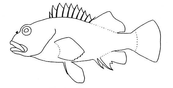
| Soft dorsal long, of 29-33 rays; color pattern of alternating oblique black and white bands. [Fig. 367] |
| Latridae |
|
|
| b |
| Soft dorsal short, of 17 or fewer rays; color pattern not of alternating oblique black and white bands. Tips of dorsal spines have one or more projecting cirri. [Fig. 368] |
| Cirrhitidae |
|
|
| |
|
52a |
| Lateral line absent. Lower jaw heavy and protruding; mouth almost vertical. Elongate with continuous dorsal of 28-66 soft rays. [Fig. 395] |
| Microdesmidae |
|
|
| b |
| Lateral line present, at least forward. |
|
53 |
| |
|
53a |
| A single, sharp, more or less conical spine on the opercle. Anal without spines and with 18 or more soft rays. |
|
54 |
| b |
| No single, sharp, more or less conical spine on the opercule. |
|
55 |
| |
|
54a |
| Caudal fin with a pair of prominent longitudinal black bars; anal fin with 46-55 rays. [Fig. 369] |
| Malacanthidae |
|
|
| b |
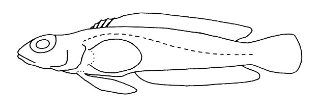
| Caudal fin without prominent longitudinal bars; anal with 14-22 rays. Eyes oriented as much dorsally as laterally. [Fig. 370] |
| Pinguipedidae |
|
|
| |
|
55a |
| Branched caudal rays about 15; gill covers attached to the isthmus far forward or entirely free from it. Two or more sharp anal spines. |
|
57 |
| b |
| Branched caudal rays 11 or 12; gill covers broadly attached to the isthmus or to one another by a membrane across the isthmus. Scales cycloid; caudal often lunate, but never forked. |
|
56 |
| |
|
56a |
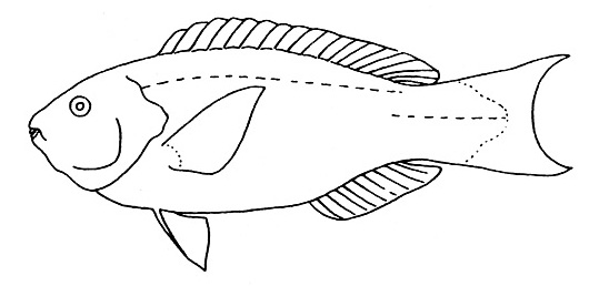
| Jaw teeth either fused into a beaklike structure or with 2 to several series of overlapping incisors in front. [Fig. 372] |
| Scaridae |
|
|
| b |
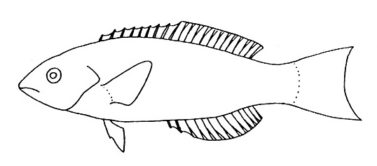
| Jaws with a single series of separate teeth in front, often canines. [Fig. 371] |
| Labridae (in part) |
|
|
| |
|
57a |
| Teeth fused into a beaklike structure. [Fig. 373] |
| Oplegnathidae |
|
|
| b |
| Teeth not fused into a beaklike structure. |
|
58 |
| |
|
58a |
| Anal fin with 2 spines. Lateral line ending under the soft dorsal fin; a single nostril on each side of the head. [Fig. 374] |
| Pomacentridae |
|
|
| b |
| Anal fin with 3 or more spines. |
|
59 |
| |
|
59a |
| Sides plain silvery, darker above. [Fig. 375] |
| Kuhliidae |
|
|
| b |
| Sides never plain silvery. |
|
60 |
| |
|
60a |
| Mouth small, not reaching the level of the anterior nostril; teeth more or less flexible, like the teeth of a comb. Body deep and strongly compressed. |
|
61 |
| b |
| Mouth moderate or large, the maxillary reaching to behind the level of the anterior nostril; teeth firm. |
|
63 |
| |
|
61a |
| Pelvic fins originating behind pectoral base. Dorsal with 11 spines and 16-18 soft rays. Body with 6 black, nearly horizontal stripes. [Fig. 377] |
| Microcanthidae |
|
|
| b |
| Pelvic fins originating below pectoral base. Dorsal not with 11 spines and 16-18 soft rays. |
|
62 |
| |
|
62a |
| Prominent spine at the corner of the preopercle. No enlarged scale at base of pelvics. [Fig. 396] |
| Pomacanthidae |
|
|
| b |
| No prominent spine at corner of preopercle. Enlarged scale at base of pelvics. [Fig. 376] |
| Chaetodontidae |
|
|
| |
|
63a |
| Anal soft rays 12 or more. Pelvic fins attached to abdomen by broad membrane. Large eyes; color primarily red, sometimes silvery. Scales small and rough. [Fig. 379] |
| Priacanthidae |
|
|
| b |
| Anal soft rays 11 or fewer. |
|
64 |
| |
|
64a |
| Posterior maxillary completely exposed when the mouth is closed. Three flat opercular spines. [Fig. 382] |
| Serranidae |
|
|
| b |
| Posterior maxillary slips under cheek when mouth is closed. Single spine on opercle, if present. |
|
65 |
| |
|
65a |
| Dorsal with 11 to 15 soft rays (rarely 10). No spine on opercle. [Fig. 381] |
| Lutjanidae |
|
|
| b |
| Dorsal with 9-10 soft rays. Single flat spine on opercle. [Fig. 380] |
| Lethrinidae |
|
|
| |
| Images |
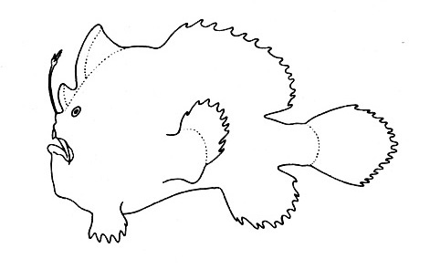 |
| Figure 330. Representative of the Antennariidae family (Gosline & Brock, 1960, fig. 23) |
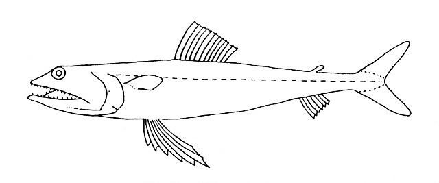 |
| Figure 331. Representative of the family Synodontidae (Gosline & Brock, 1960, fig. 32) |
 |
| Figure 332. Representative of the family Aulostomidae (Gosline & Brock, 1960, fig. 41) |
 |
| Figure 333. Representative of the family Fistulariidae (Gosline & Brock, 1960, fig. 42) |
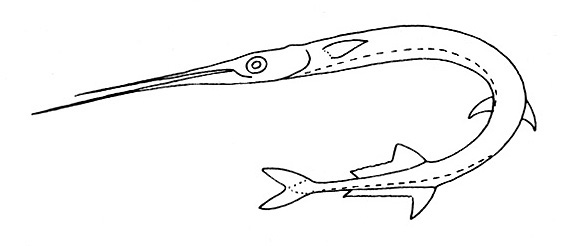 |
| Figure 334. Representative of the family Belonidae (Gosline & Brock, 1960, fig. 47) |
 |
| Figure 335. Representative of the family Hemiramphidae (Gosline & Brock, 1960, fig. 48) |
 |
| Figure 336. Representative of the family Elopidae (Gosline & Brock, 1960, fig. 51) |
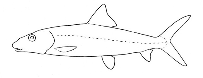 |
| Figure 337. Representative of the family Albulidae (Gosline & Brock, 1960, fig. 52) |
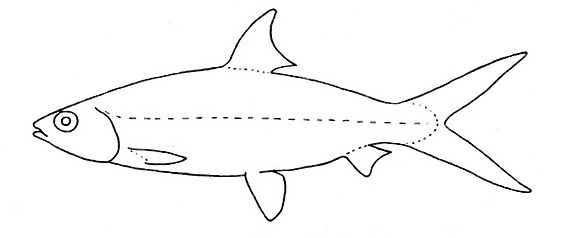 |
| Figure 338. Representative of the family Chanidae (Gosline & Brock, 1960, fig. 53) |
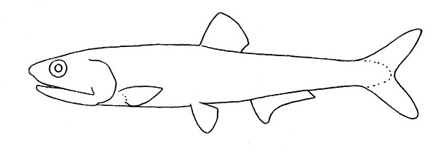 |
| Figure 339. Representative of the family Engraulidae (Gosline & Brock, 1960, fig. 54) |
 |
| Figure 340. Representative of the family Clupeidae (Gosline & Brock, 1960, fig. 55) |
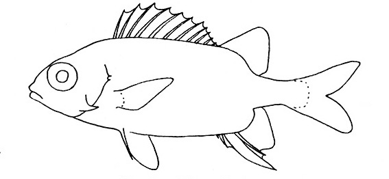 |
| Figure 341. Representative of the family Holocentridae (Gosline & Brock, 1960, fig. 59) |
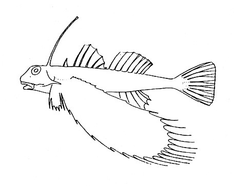 |
| Figure 342. Representative of the family Dactylopteridae (Gosline & Brock, 1960, fig. 63) |
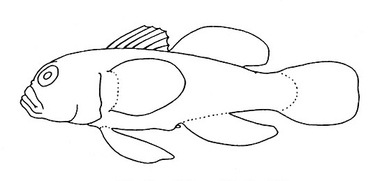 |
| Figure 343. Representative of the family Gobiidae (Gosline & Brock, 1960, fig. 65) |
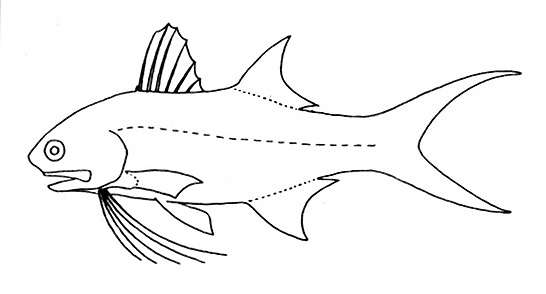 |
| Figure 344. Representative of the family Polynemidae (Gosline & Brock, 1960, fig. 70) |
 |
| Figure 345. Representative of the family Tripterygiidae (Gosline & Brock, 1960, fig. 75) |
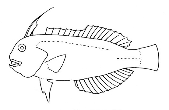 |
| Figure 346. Representative of the family Labridae (Gosline & Brock, 1960, fig. 76) |
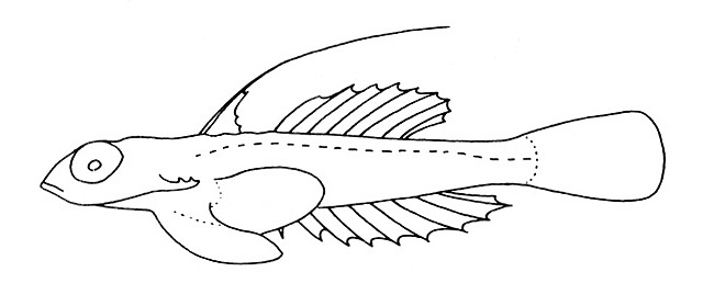 |
| Figure 347. Representative of the family Callionymidae (Gosline & Brock, 1960, fig. 77) |
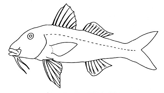 |
| Figure 348. Representative of the family Mullidae (Gosline & Brock, 1960, fig. 79) |
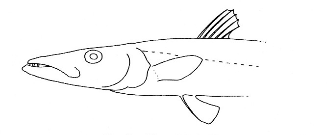 |
| Figure 349. Representative of the family Sphyraenidae (Gosline & Brock, 1960, fig. 81) |
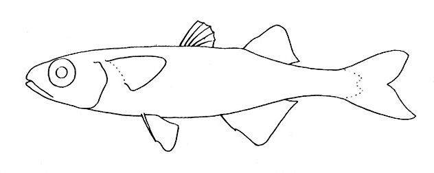 |
| Figure 350. Representative of the family Atherinidae (Gosline & Brock, 1960, fig. 82) |
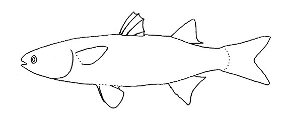 |
| Figure 351. Representative of the family Mugilidae (Gosline & Brock, 1960, fig. 83) |
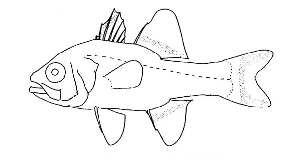 |
| Figure 352. Representative of the family Apogonidae (Gosline & Brock, 1960, fig. 84) |
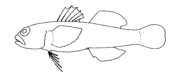 |
| Figure 353. Representative of the family Gobiidae (Gosline & Brock, 1960, fig. 85) |
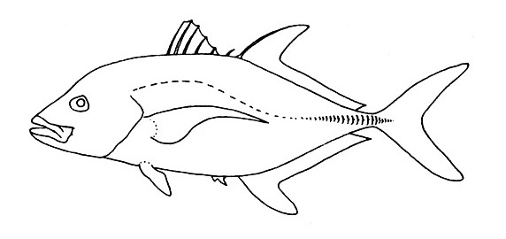 |
| Figure 354. Representative of the family Carangidae (Gosline & Brock, 1960, fig. 89) |
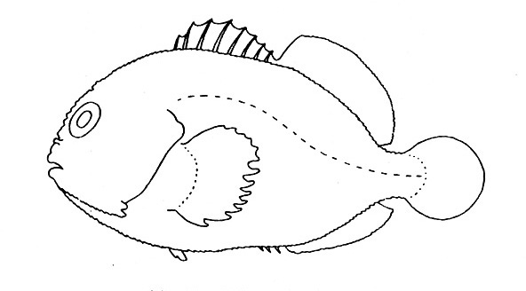 |
| Figure 355. Representative of the family Caracanthidae (Gosline & Brock, 1960, fig. 90) |
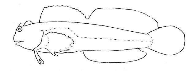 |
| Figure 356. Representative of the family Blenniidae (Gosline & Brock, 1960, fig. 93) |
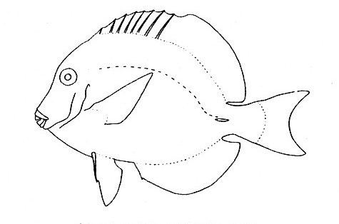 |
| Figure 358. Representative of the family Acanthuridae (Gosline & Brock, 1960, fig. 97) |
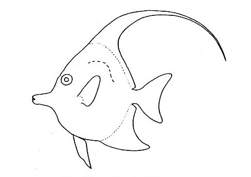 |
| Figure 359. Representative of the family Zanclidae (Gosline & Brock, 1960, fig. 98) |
 |
| Figure 361. Representative of the family Samaridae (Gosline & Brock, 1960, fig. 20) |
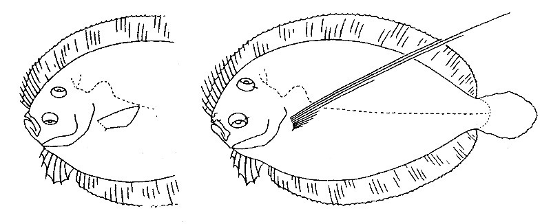 |
| Figure 362. Representative of the family Bothidae; female left, male right (Gosline & Brock, 1960, fig. 21) |
 |
| Figure 363. Representative of the family Ophidiidae (Gosline and Brock, 1960, fig. 95) |
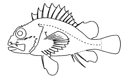 |
| Figure 364. Representative of the Scorpaenidae family (Gosline & Brock, 1960, fig. 100) |
 |
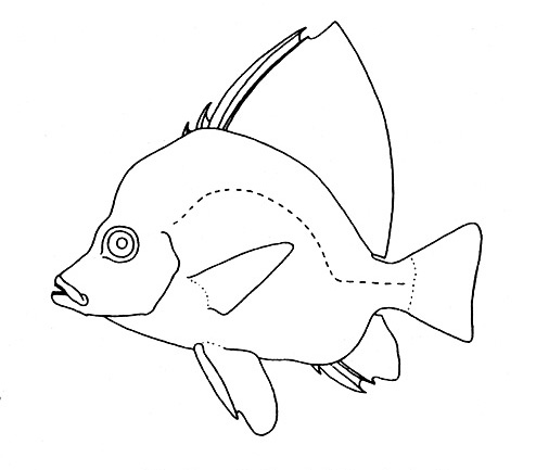
| Figure 365. Representative of the Echeneidae family (Gosline & Brock, 1960, fig. 101) |
 |
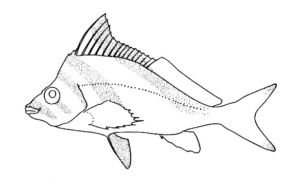
| Figure 366. Representative of the Pentacerotidae family (Gosline & Brock, 1960, fig. 103) |
 |
| Figure 367. Representative of the Latridae family (Gosline & Brock, 1960, fig. 105) |
 |
| Figure 368. Representative of the Cirrhitidae family (Gosline & Brock, 1960, fig. 106) |
 |
| Figure 369. Representative of the Malacanthidae family (Gosline & Brock, 1960, fig. 109) |
 |
| Figure 370. Representative of the Pinguipedidae family (Gosline & Brock, 1960, fig. 110) |
 |
| Figure 371. Representative of the Labridae family (Gosline & Brock, 1960, fig. 112) |
 |
| Figure 372. Representative of the Scaridae family (Gosline & Brock, 1960, fig. 113) |
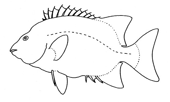 |
| Figure 373. Representative of the Oplegnathidae family (Gosline & Brock, 1960, fig. 114) |
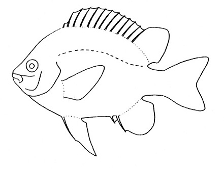 |
| Figure 374. Representative of the Pomacentridae family (Gosline & Brock, 1960, fig. 115) |
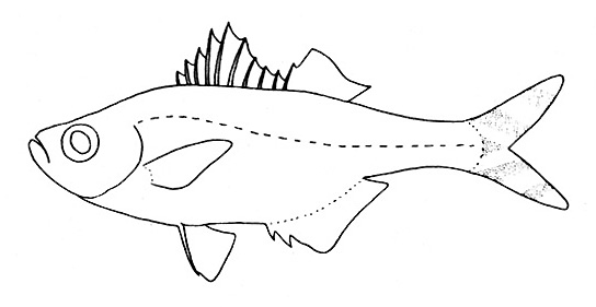 |
| Figure 375. Representative of the Kuhliidae family (Gosline & Brock, 1960, fig. 116) |
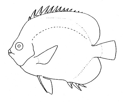 |
| Figure 376. Representative of the Chaetodontidae family (Gosline & Brock, 1960, fig. 117) |
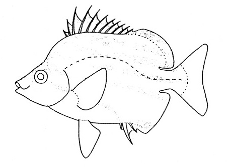 |
| Figure 377. Representative of the Microcanthidae family (Gosline & Brock, 1960, fig. 118) |
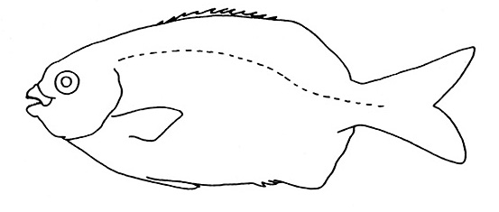 |
| Figure 378. Representative of the Kyphosidae family (Gosline & Brock, 1960, fig. 119) |
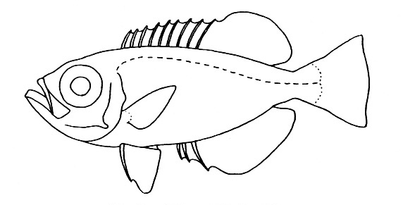 |
| Figure 379. Representative of the Priacanthidae family (Gosline & Brock, 1960, fig. 121) |
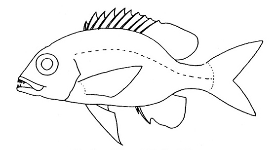 |
| Figure 380. Representative of the Lethrinidae family (Gosline & Brock, 1960, fig. 122) |
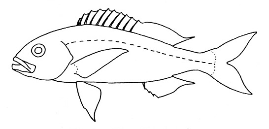 |
| Figure 381. Representative of the Lutjanidae family (Gosline & Brock, 1960, fig.123) |
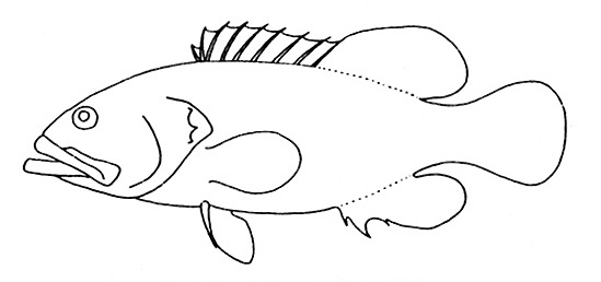 |
| Figure 382. Representative of the Serranidae family (Gosline & Brock, 1960, fig. 124) |
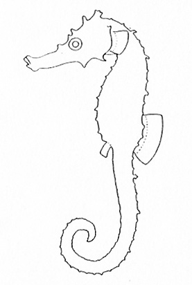 |
| Figure 383. Representative of the Syngnathidae family (Gosline & Brock, 1960, fig. 125) |
 |
| Figure 384. Representative of the Congridae family (Gosline & Brock, 1960, fig. 131) |
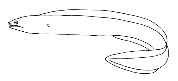 |
| Figure 385. Representative of the Muraenidae family (Gosline & Brock, 1960, fig. 135) |
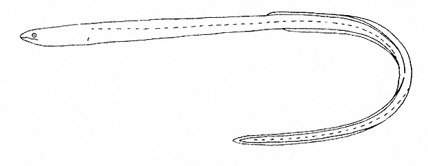 |
| Figure 386. Representative of the Ophichthidae family (Gosline & Brock, 1960, fig. 136) |
 |
| Figure 387. Representative of the Syngnathidae family (Gosline & Brock, 1960, fig. 138) |
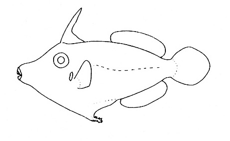 |
| Figure 388. Representative of the Monacanthidae family (Gosline & Brock, 1960, fig. 139) |
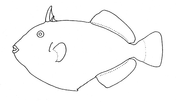 |
| Figure 389. Representative of the Balistidae family (Gosline & Brock, 1960, fig. 140) |
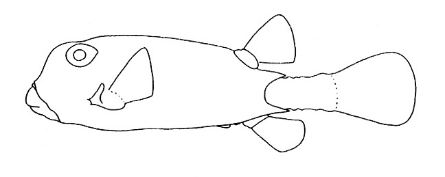 |
| Figure 390. Representative of the Ostraciidae family (Gosline & Brock, 1960, fig. 141) |
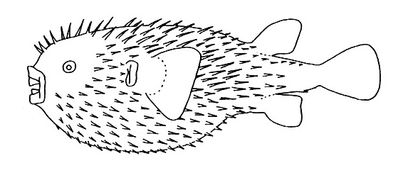 |
| Figure 391. Representative of the Diodontidae family (Gosline & Brock, 1960, fig. 142) |
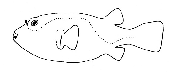 |
| Figure 392. Representative of the Tertaodontidae family (Gosline & Brock, 1960, fig. 143) |
 |
| Figure 393. Representative of the Ammodytidae family (Gosline & Brock, 1960, fig. 146) |
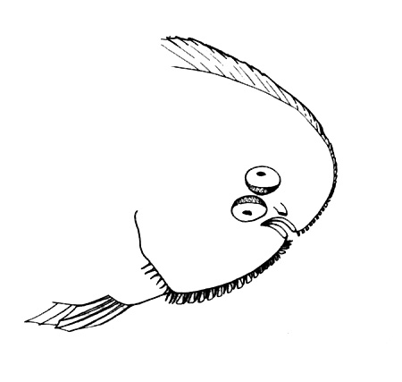 |
| Figure 394. Representative of the family Soleidae (Gosline & Brock, 1960, fig. 177) |
 |
| Figure 395. Representative of the family Microdesmidae (Nelson, 2006, pg. 423) |
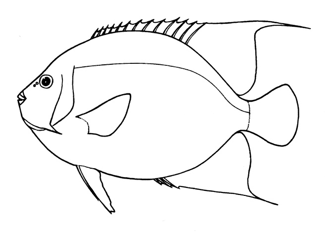 |
| Figure 396. Representative of the family Pomacanthidae (Nelson, 2006, pg. 379) |
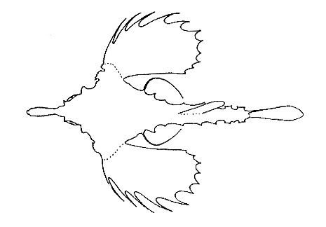 |
| Figure 397. Representative of the family Pegasidae (Gosline & Brock, 1960, fig. 92) |


