Key (online version)
|
|
1a |
| Wings normally developed. |
|
2 |
| b |
| Wings reduced to a minute vestigial lobe, halteres present. Whole surface of first abdominal tergite of male with a distinctive granulated ommatidia-like pattern. |
| Thripomorpha paludicola (Enderlein, 1905) |
|
|
| |
|
2a |
| Scutum with an elevated U-shaped ridge [Fig. 749]. Anterior tibia with a strongly developed apical spur. Costa strongly swollen at junction with R4+5 [Fig. 750]. |
| Aspistinae |
|
3 |
| b |
| Scutum without an elevated ridge, rounded or flattened in lateral view. Fore tibia not produced into an apical spur. Costa not swollen at junction with R4+5. |
|
5 |
| |
|
3a |
| Flagellum of antenna 5-segmented in both sexes. Fore femur simple, unarmed beneath (Holarctic, boreal). |
| Arthria analis (Kirby, 1837) |
|
|
| b |
Flagellum of antenna 6-segmented in female, 8-10-segmented in male. Fore femur with spines beneath.
Three species in Fauna Europaea. |
| Aspistes Meigen, 1818 |
|
4 |
| |
|
4a |
|
| Aspistes berolinensis Meigen, 1818 |
|
|
| b |
|
| Aspistes helleni Krivosheina, 2000 |
|
|
| |
|
5a |
R4+5 and M fused for a distance, cross-vein R-M obliterated, stem of M fork arising from R4+5, well distally from transverse vein (base of R4+5) [Fig. 753]. Hind tibia flattened, strongly swollen in apical third. A pseudotrachea on labella.
Four species in Fauna Europaea. |
| Ectaetiinae: Ectaetia Enderlein, 1912 |
|
|
| b |
| Cross-vein R-M present, even if generally very short, often oblique or even horizontal, R4+5 and M separated, touching at most in one point [Fig. 751] [Fig. 752] and base of M fork arising from just before or below transverse vein (base of R4+5). Hind tibia normal, at most slightly enlarged apically. Labella devoid of pseudotrachea. |
|
6 |
| |
|
6a |
| No false vein visible between M2 and CuA1 [Fig. 751]. Posterior veins and membrane with macrosetae among microtrichia. M fork incomplete, base of M1 lacking [Fig. 751]. Sperm pump of male directly attached to genital capsule. |
| Psectrosciarinae |
|
7 |
| b |
| False vein present as a concave fold between M2 and CuA1, sometimes bifurcated at apex [Fig. 752] (this fold is not always very visible, the wing must be thoroughly observed under different angles of view; usually the fold appears clearly over a dark surface). Posterior veins and membrane with or without macrosetae among the microtrichia. M fork generally complete (except in Thripomorpha with well-visible false vein present). Sperm-pump of male attached to genitalia only by an elongated sperm-duct, lying apparently free in abdomen. |
| Scatopsinae |
|
19 |
| |
|
7a |
Body strongly compressed laterally, elongated. R4+5 joining the costa gently in a smooth curve [Fig. 754].
Two species in Fauna Europaea. |
| Psectrosciara Kieffer in Enderlein, 1911 |
|
|
| b |
Body not strongly compressed laterally nor elongated. R4+5 joining rather abruptly the costa, forming with it an angle vicinous to 90 degrees [Fig. 755].
Twenty four species in Fauna Europaea, one described later from Central Europe by Haenni & Martinovský in 2014. |
| Anapausis Enderlein, 1912 |
|
8 |
| |
|
8a |
| Vein M1 bent anteriorly at wing margin; macrotrichia on wing membrane anterior to M1. Male tergite 8 with two parallel strap-like curved arms, subapically joined by median bridge. Female sternite 7 with broad lateral lobes widely separated medially. |
| Anapausis talpae (Verrall, 1912) |
|
|
| b |
| Vein M1 not bent anteriorly at wing margin; macrotrichia mainly confined to membrane behind M1. Male tergite 8 without apically fused lateral arms. Female sternite 7 not divided into two lobes. |
|
9 |
| |
|
9a |
| First tarsomeres without comb-like rows of stout setae. Male tergite 8 rounded apically with small median indentation, lacking paired processes. Female tergite 8 with small projection medially; tergite 9 absent. |
| Anapausis nigripes (Zetterstedt, 1860) |
|
|
| b |
| First tarsomeres with comb-like rows of spiniform setae. Male tergite 8 with a pair of posteroventrally directed tapered blunt processes. Female tergite 8 without medial projection; tergite 9 well developed. |
|
|
| |
|
10a |
|
11 |
| b |
|
15 |
| |
|
11a |
| Sternite 6 broadly emarginate medially, lateral parts simple and not bilobed. Tergite 8 with pair of processes short, not longer than gap between them [Fig. 315]. |
| Anapausis rectinervis Duda, 1928 |
|
|
| b |
| Sternite 6 deeply emarginate medially, lateral part not simple but variously lobed. |
|
12 |
| |
|
12a |
| Sternite 6 with lateral part not bilobed medially, but with apical margin angled internally and bearing a posteriorly directed thumb-like process near this angle. Tergite 8 with pair of processes about as long or a little longer than gap between them [Fig. 316]. |
| Anapausis pollicata Chandler, 1999 |
|
|
| b |
| Sternite 6 with lateral part deeply bilobed medially. Tergite 8 with pair of processes longer than gap between them. |
|
13 |
| |
|
13a |
| Sternite 6 with narrow emargination, each lateral part with anterior margin evenly curved and posterior lobe pointed apically [Fig. 317]. Stemite 5 with large rounded anterior lobes, broader than the gap between them [Fig. 317]. |
| Anapausis dalmatina Duda, 1928 |
|
|
| b |
| Sternite 6 with wider emargination, each lateral part with anterior margin concave on inner part and posterior lobe not pointed apically. Stemite 5 with rounded anterior lobes small and widely separated. |
|
14 |
| |
|
14a |
| Sternite 6 with posterior lobes only slightly indented subapically; tergite 8 dark brown, with processes shorter and more slender [Fig. 318]. |
| Anapausis soluta (Loew, 1846) |
|
|
| b |
| Sternite 6 with posterior lobes strongly indented subapically to give a hooked appearance; tergite 8 usually more reddish with processes longer and thicker [Fig. 319]. |
| Anapausis floricola Chandler, 1999 |
|
|
| |
|
15a |
| Tergite 9 distinctly longer than broad, with straight basal margin and deeply inset in tergite 8 which is short medially. Stemite 8 emarginate apically. |
| Anapausis rectinervis Duda, 1928 |
|
|
| b |
| Tergite 9 not longer than broad, with curved basal margin. Tergite 8 broader medially with curved apical margin. |
|
16 |
| |
|
16a |
| Stemite 9 produced apically to a point, protruding beyond tergite 9. Furca with a pair of diverging (ventrally directed) anterior processes [Fig. 757]. |
| Anapausis sp. indet. 1 |
|
|
| b |
| Stemite 9 rounded apically. |
|
17 |
| |
|
17a |
| Tergite 9 more angular and deeply inset in concave apical margin of tergite 8. Furca with single slender anterior process [Fig. 758]. |
| Anapausis dalmatina Duda, 1928 |
|
|
| b |
| Tergite 9 broadly ovate, more shallowly inset in tergite 8 [Fig. 759] [Fig. 760]. |
|
18 |
| |
|
18a |
| Tergite 8 without a rounded lobe laterally. Furca with a pair of short diverging (sometimes anteroventrally directed) anterior processes [Fig. 760]. |
| Anapausis soluta (Loew, 1846) |
|
|
| b |
| Tergite 8 with bluntly rounded lateral lobe (extended more distally than in dalmalina). Furca with single elongate anterior process [Fig. 759]. |
| Anapausis floricola Chandler, 1999 |
|
|
| |
|
19a |
Presence of macrosetae on posterior veins, and also on membrane in some genera [Fig. 763]. Stem of haltere without setae.
(In minute Parascatopse, at least 12 macrosetae present on CuA2, sometimes difficult to see; high magnification necessary). |
| Rhegmoclematini |
|
20 |
| b |
| No macrosetae on posterior veins and on membrane [Fig. 761]. Stem of haltere with setae in Palaearctic genera (except in extra-limital Lumpuria with quadrate scutum, dichoptic eyes in both sexes and elongated hind tibia). |
|
25 |
| |
|
20a |
Presence of macrosetae on M1, M2, or both. M1 incomplete basally in Palaearctic species [Fig. 762]. Flagellum of antenna 10-segmented. First abdominal tergite of male often with a granulated ommatidia-like structure.
Nine species in Fauna Europaea:
bifida (Zilahi-Sebess, 1956) (= collini (Cook, 1969))
cooki (Hutson, 1970)
coxendix (Verrall, 1912)
freyi (Cook, 1969)
haennii Martinovsky, 1996
halteratum (Meigen, 1838)
paludicola Enderlein, 1905 (= edwardsi (Collin, 1954))
verralli (Edwards, 1934)
vockerothi (Cook, 1969) |
| Thripomorpha Enderlein, 1905 |
|
|
| b |
| No macrosetae on M1 and M2, macrosetae present only on CuA2 vein. M fork complete [Fig. 763]. |
|
21 |
| |
|
21a |
| Flagellum of antenna 10-segmented. |
| Neorhegmoclemina Cook, 1955: Neorhegmoclemina catharinae Haenni, 1997 |
|
|
| b |
| Flagellum of antenna 8-segmented. |
|
22 |
| |
|
22a |
Male with sternite 7 shield-shaped [Fig. 424]. A regular distinct supraalar row of setae. Spiracular sclerite elongated, triangular.
Four species in Fauna Europaea. |
| Rhegmoclemina Enderlein, 1936 |
|
|
| b |
| Male sternite 7 not shield-shaped. No distinct supraalar row of setae. Spiracular sclerite not elongated, rounded in general shape. Size very small, wing length about 1 mm (mostly coastal or halophilous). |
| Parascatopse Cook, 1955 |
|
23 |
| |
|
23a |
| Legs with tibiae broadly whitish at base, tarsi and halteres pale brown. |
| Parascatopse minutissima (Verrall, 1886) |
|
|
| b |
| Legs uniformly darkish, though not as dark as body. Halteres at least darkened on knob. |
|
24 |
| |
|
24a |
| Sternite 7 broadly triangular. Lateral appendages in the genital capsule wide, rounded apically. |
| Parascatopse litorea (Edwards, 1925) |
|
|
| b |
| Sternite 7 with median posterior projection truncate apically [Fig. 764]. Lateral appendages in the genital capsule narrow, contorted. |
| Parascatopse distorta Haenni & Pollini, 2015 |
|
|
| |
|
25a |
| Thorax stout, as wide as long, quadrate or nearly so. Wings densely microtrichiose. |
| Colobostematini |
|
26 |
| b |
| Thorax narrow, much longer than wide. |
|
32 |
| |
|
26a |
A complete supernumerary cross-vein between M1 and R4+5 [Fig. 765].
Four species in Fauna Europaea. |
| Holoplagia Enderlein, 1912 |
|
27 |
| b |
| No complete supernumerary cross-vein between M1 and R4+5 [Fig. 766], but M1 often angled at 1/4 basal, sometimes with a stump of vein directed towards R4+5 [Fig. 647]. |
|
31 |
| |
|
27a |
|
|
| b |
|
28 |
| |
|
28a |
| Wing without bulla or microtichose patch between veins M1 and M2. |
|
29 |
| b |
| Wing with bulla or microtichose patch between veins M1 and M2. |
|
30 |
| |
|
29a |
| Wings with CuA1 slightly bent at apex (compare with [Fig. 641], but not as stong in this figure); M1 and M2 parallel and both slightly bent forwards. |
| Holoplagia transversalis (Loew, 1846) |
|
|
| b |
| Wings with CuA1 straight; M1 and M2 not curved basally but parallel and curved forwards along their length, slightly divergent apically. |
| Holoplagia richardsi Freeman, 1985 |
|
|
| |
|
30a |
| Genitalia |
| Holoplagia bullata (Edwards, 1925) |
|
|
| b |
Genitalia
|
| Holoplagia lucifaga (Loew,1870) |
|
|
| |
|
31a |
Flagellar segments with a distinct neck, short, bead-like or conical. Occiput regularly rounded posteriorly. Eyes dichoptic in female, hardly touching and nearly dichoptic in male. Thorax and abdomen dull, not shiny.
Eleven species keyed by Haenni in 2013 but he mentions at least 18 Western Palaearctic species with probably more undescribed Mediterranean species. |
| Colobostema Enderlein, 1926 |
|
|
| b |
Flagellar segments devoid of neck, cylindrical and more or less elongated. Occiput hollowed out posteriorly, head short. Eyes clearly dichoptic in both sexes. Posterior tibia elongated.
Not yet recorded in the Palaearctic, 5 Oriental spp., Nepal; Haenni 1988b. |
| Lumpuria Edwards, 1928 |
|
|
| |
|
32a |
Wings infuscate, dull. A cluster of spiniform setae on sternite 7 posteriorly, beside usual setae (visible at high magnification). R4+5 long, joining costa at about 3/4 of wing length.
Two species in Fauna Europaea. |
| Ferneiella Cook, 1977 |
|
|
| b |
| Wings clear, more or less hyaline or milky, at most slightly infuscated. No spiniform setae on posterior margin of sternite 7. R4+5 of variable length. |
|
33 |
| |
|
33a |
A complete supernumerary cross-vein between M1 and R4+5 (though translucent, this vein is
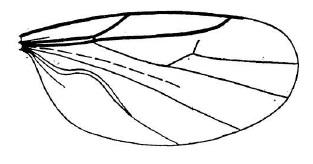
well-visible, especially under tangential light). Vein CuA2 strongly recurved towards wing base [Fig. 641]. Tarsi white, contrasting with rest of body and legs. |
| Efcookella Haenni, 1998: Efcookella albitarsis (Zetterstedt, 1850) |
|
|
| b |
| No complete supernumerary cross-vein between M1 and M fork evenly curved or smoothly angled at 1/4 basal, with at most one anteriorly directed stump of vein arising towards R4+5. |
|
34 |
| |
|
34a |
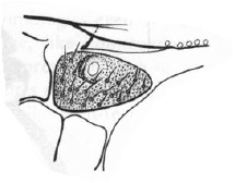
| Second costal section practically equal to first section, or longer. R4+5 long, reaching costa at 2/3 of wing length or further [Fig. 761]; if hardly so (some Reichertella), then CuA2 smoothly curved towards wing tip [Fig. 761], not reaching abruptly hind margin of wing. Anterior spiracular sclerite nearly as high as long, with a comparatively large spiracular opening [Fig. 767]. Palpus rounded, oval or truncate at apex, usually shorter or approximately as long as labella. |
| Scatopsini |
|
35 |
| b |
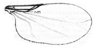
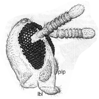
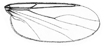
Second costal section clearly shorter than first section. R4+5 short [Fig. 648] [Fig. 649], reaching costa at middle of wing length or at most somewhat further, and then CuA2 reaching hind margin of wing abruptly, at a nearly right angle [Fig. 648]. Palpus usually large, sausage shaped or pointed
at apex [Fig. 769], usually at least as long as labella or longer; anterior spiracular sclerite longer than high, spiracular opening comparatively smaller [Fig. 768]. Thorax and abdomen entirely covered with a vestiture of minute microtrichia beside the usual pilosity (high magnification required). |
| Swammerdemellini |
|
41 |
| |
|
35a |
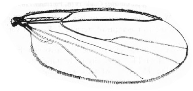
| Fork of M with an angle at 1/4-1/3 basal of M1 and an anteriorly directed stump of vein arising from this angle (translucent and sometimes weakly visible) [Fig. 647]; if no stump of vein visible (Pharsoreichertella hamifera (Strobl)), then M fork strongly assymetrical, with M1 strongly bent and M2 nearly straight [Fig. 770]. |
|
36 |
| b |
| Fork of M practically symmetrical, with M1 and M2 evenly and smoothly curved, without any indication of an angle or a stump of vein at 114-113 basal [Fig. 771]. |
|
39 |
| |
|
36a |
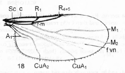
Tarsomere 1 of male hind leg shorter than tarsomere 2, or equal in length, armed with spines. Tergite 7 of male not projecting posteriorly, concealed under tergite 6 and weakly sclerotized. M1 with a well-marked angle at 1/4 basal and a stump of vein directed towards R4+5, well-visible though translucent [Fig. 647]. Abdominal tergites and sternites bright, devoid of microtrichia beside the usual pilosity.
Three species in Fauna Europaea. |
| Scatopse Geoffroy, 1762 |
|
37 |
| b |
Tarsomere 1 of hind leg unmodified in male, longer than tarsomere 2. Tergite 7 of male well-visible and sclerotized, projecting posteriorly in 1 or 2 lobes. Angle of M1 not very marked, with a weakly visible anteriorly directed stump or, if stump absent, then fork of M strongly asymmetrical [Fig. 770]. Abdominal tergites and sternites dull, at most subshiny, with a vestiture of microtrichia (visible at high magnification) beside the usual pilosity.
Three species in Fauna Europaea. |
| Pharsoreichertella Cook, 1974 |
|
|
| |
|
37a |
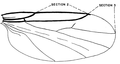
| Entire length of vein R4+5 ventrally with 3-5 rows of setulae; section 2 of C at least 1.25 times as long as section 3 [Fig. 647]. Male: penis valves apically pointed or nearly so, claw-like [Fig. 644]. Female: sternum 8 with a pair of valvifers [Fig. 646]. |
| Scatopse notata (Linnaeus, 1758) |
|
|
| b |
| Vein R4+5 ventrally with 1-3 rows of setulae, at least on its proximal part; section 2 of C equal to section 3 or shorter [Fig. 645]. Male: penis valves apically broadly rounded. Female: sternum 8 without valvifers. |
|
38 |
| |
|
38a |
Distal third of vein R4+5 ventrally with 2-3 irregular rows of setulea. Male with first segment of both mid and hind tarsi apically produced, bearing a cluster of spines; gonocoxites broad, rounded, with longest axis nearly transverse to axis of hypopygium; penis short, blunt apically [Fig. 773]. Female with lobes of sternum 8 broad with inner margin straight or nearly so [Fig. 773]; cerci large, proximally largely overlapped by tergum 8 [Fig. 774].
[Fig. 772] |
| Scatopse globulicauda Laštovka & Haenni, 1981 |
|
|
| b |
| Entire length of vein R4+5 ventrally with a single row of sotulae. Male with first segment of hind tarsus only produced and bearing spines apically; gonocoxites slender, with axis nearly parallel to axis of hypopygium; penis long, pointed apically. Female with lobes of sternum 8 narrow with inner margin sinuous; cerci small, not proximally overlapped by tergum 8. |
| Scatopse lapponica Duda, 1928 |
|
|
| |
|
39a |
Abdominal sternites (and also tergites) bright, devoid of microtrichia beside the usual pilosity. R4+5 long, reaching the costa in a smooth curve well beyond 2/3 of wing length [Fig. 771]. Male pregenital segment (sternite and sometimes also tergite 7) diversely modified, with more or less developed, often asymmetrical lateral posterior projections. Penis elongated, filamentous, 2-3 branched, diversely modified at apex; 1-2 pairs of hypogynial valves in female.
Thirteen species in Fauna Europaea. |
| Apiloscatopse Cook, 1974 |
|
|
| b |
Abdominal sternites dull or at most subshiny, with a vestiture of microtrichia (visible at high magnification) beside the usual pilosity. R4+5 shorter, reaching the costa at about 2/3 of wing length, or if longer (Reichertella geniculata), then running parallel to the costa and joining this vein after a marked knee. Male tergite 7 partly desclerotized or reduced, much shorter than sternite 7. Penis shorter and thick, more or less contorted, enlarged or flattened. No hypogynial valves in female.
Three species in Fauna Europaea. |
| Reichertella Enderlein, 1912 |
|
40 |
| |
|
40a |
| Vein R4+5 parallel to costa for most of its length, fairly sharply turned towards costa at its end [Fig. 434], |
| Reichertella geniculata (Zetterstedt, 1850) |
|
|
| b |
| Vein R4+5 more or less converging gradually on to costa, not sharply turned towards it apically, costa shorter, anal angle more obtuse [Fig. 435]. |
| other Reichertella sp. |
|
|
| |
|
41a |
M fork short or very short, always shorter than M stem [Fig. 415]. R1 and R4+5 joining costa very close to each other; distance between them (second costal section) shorter than length of R1 from origin of R4+5 to costa [Fig. 415]. Palpus pointed apically [Fig. 769].
Six species in Fauna Europaea, one species described later by Haenni from Spain in 2015. |
| Swammerdamella Enderlein, 1912 |
|
42 |
| b |
| M fork obviously longer than stem [Fig. 648]. |
|
43 |
| |
|
42a |
Flagellum 8-segmented. M-fork longer, branches almost as long as or longer than stem.
Swammerdamella pediculata (Duda, 1928), Swammerdamella genypodis Cook, 1972, Swammerdamella spinigera Haenni, 2009 |
|
|
| b |
Flagellum 7-segmented. M-fork very short, branches much shorter than stem.
Swammerdamella acuta Cook, 1956: Thorax relatively narrow; anterior spiracle below the midline of the spiracular plate; male tergite 6 with angular posterior margin.
Swammerdamella adercotris Cook, 1972: Thorax relatively broad; anterior spiracle on the midline of the spiracular plate; male tergite 6 shorter with more blunt, bar-like projection on posterior margin
Swammerdamella brevicornis (Meigen, 1830): Male tergite 6 longer with angular distinctly more produced, pointed
[i]Swammerdammella jindrichi Haenni in Haenni & Roháček, 2020 |
|
|
| |
|
43a |
| Anterior spiracular sclerite a large, rather quadrate plate, twice as long as high. Male tergite 7 asymmetrically produced posteriorly as a narrow spatulate process. Aedeagus elongated, coiled. Female sternite 8 with a pair of conspicuous, fingerlike lobes projecting posteriorly. Sternites 2 and 3 sclerotized. |
| Coboldia Melander, 1916: Coboldia fuscipes (Meigen, 1830) |
|
|
| b |
| Anterior spiracular sclerite triangular elongated [Fig. 768]. Male tergite 7 symmetrical, either narrowly produced medially or not posteriorly. |
|
44 |
| |
|
44a |
Male terminalia dorsoventrally compressed. Female tergite 8 and sternite 8 without deep medial incision.
Nine species in Fauna Europaea. |
| Rhexoza Enderlein, 1936 |
|
|
| b |
| Male terminalia laterally compressed. |
|
45 |
| |
|
45a |
| Sternite 4 produced. Tergite 9 produced ventrally as a beak–like process. Posterior extension of aedeagus (“aedeagal plate”) cylindrical; parameres large basally. Female tergite 8 medially divided, sternite 8 not. |
| Quateiella Cook, 1975: Quateiella inexpectata Haenni, 1988 |
|
|
| b |
| Sternite 4 not produced. Tergite 9 without beak–like ventral extension. Aedeagal plate rather flattened; sperm duct opening on extension of aedeagus; gonostylus absent; base of parameres displaced distally. Female with tergite 8 deeply incised medially, sternite 8 broadly divided in two basally broad, apically pointed lateral lobes. |
| Cooka Amorim, 2007: Cooka incisa (Cook, 1956) |
|
|
| |
| Images |
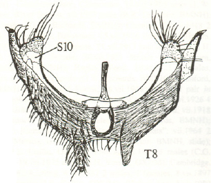 |
| Figure 315. Anapausis rectinervis, male tergite 8 and sternite 10 (after Chandler, 1999). |
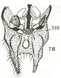 |
| Figure 316. Anapausis pollicata, male tergite 8 and sternite 10 (after Chandler, 1999). |
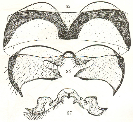 |
| Figure 317. Anapausis dalmatina, male sternites 5-7 (after Chandler, 1999). |
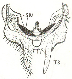 |
| Figure 318. Anapausis soluta, male tergite 8 and sternite 10 (after Chandler, 1999). |
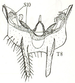 |
| Figure 319. Anapausis floricola, male tergite 8 and sternite 10 (after Chandler, 1999). |
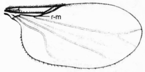 |
| Figure 415. Swammerdamella obtusa, wing (after Haenni, 1997). |
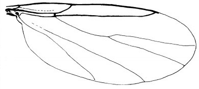 |
| Figure 416. Apiloscatopse subgracilis, male wing (after Haenni & Greve, 1995). |
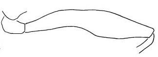 |
| Figure 417. Apiloscatopse subgracilis, male left hind femur (after Haenni & Greve, 1995). |
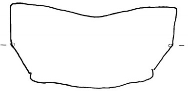 |
| Figure 418. Apiloscatopse subgracilis, male tergite 7 (after Haenni & Greve, 1995). |
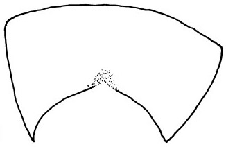 |
| Figure 419. Apiloscatopse subgracilis, male sternite 7 (after Haenni & Greve, 1995). |
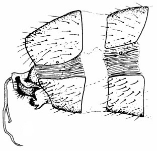 |
| Figure 420. Apiloscatopse subgracilis, male genitalia in lateral view (after Haenni & Greve, 1995). |
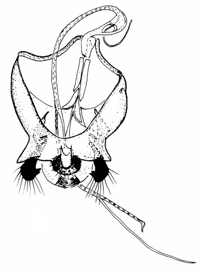 |
| Figure 421. Apiloscatopse subgracilis, male genital capsule in dorsal view (after Haenni & Greve, 1995). |
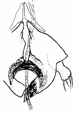 |
| Figure 422. Apiloscatopse subgracilis, male penis valves in ventral view (after Haenni & Greve, 1995). |
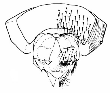 |
| Figure 423. Apiloscatopse subgracilis, female genitalia in ventral view (after Haenni & Greve, 1995). |
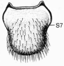 |
| Figure 424. Rhegmoclemina bimaculata, male, sternite 7 in ventral view (after Haenni, 1997). |
 |
| Figure 434. Reichertella geniculata, wing (after Freeman, 1985). |
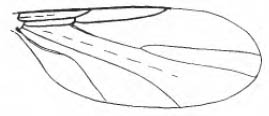 |
| Figure 435. Reichertella pulicaria, wing (after Freeman, 1985). |
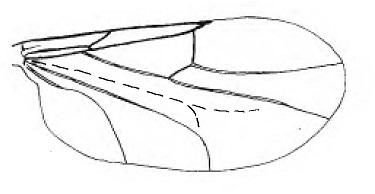 |
| Figure 641. Efcookella albitarsis, wing |
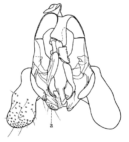 |
| Figure 644. Scatopse notata, male genital capsule in dorsal view |
 |
| Figure 645. Scatopse globulicauda, wing |
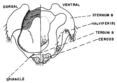 |
| Figure 646. Scatopse notata, female genitalia |
 |
| Figure 647. Scatopse notata, wing |
 |
| Figure 648. Coboldia fuscipes, wing |
 |
| Figure 649. Swammerdamella obtusa, wing |
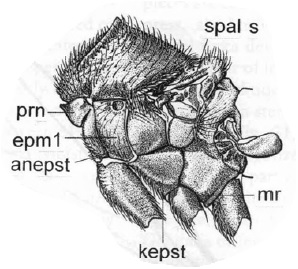 |
| Figure 749. Aspistes sp., thorax in lateral view |
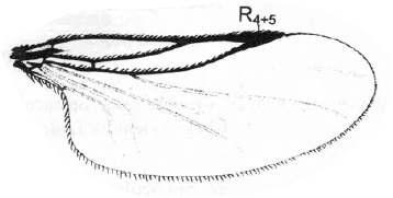 |
| Figure 750. Aspistes sp., wing |
 |
| Figure 751. Psectrosciarinae, wings |
 |
| Figure 752. Scatopsinae, wings |
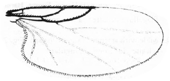 |
| Figure 753. Ectaetia sp., wing |
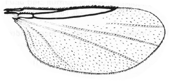 |
| Figure 754. Psectrosciara sp., wing |
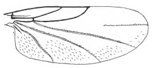 |
| Figure 755. Anapausis soluta, wing |
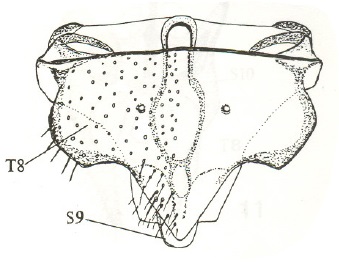 |
| Figure 756. Anapausis sp. Austria, female genitalia in dorsal view |
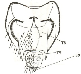 |
| Figure 757. Anapausis sp. Scotland, female genitalia in dorsal view |
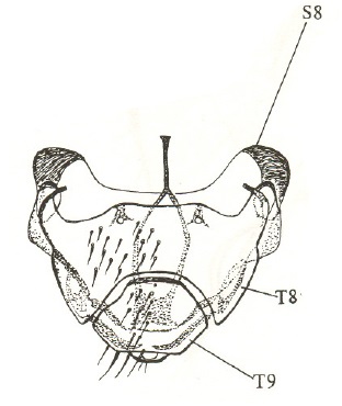 |
| Figure 758. Anapausis dalmatina, female genitalia in dorsal view |
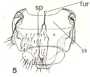 |
| Figure 759. Anapausis floricola, female genitalia in dorsal view |
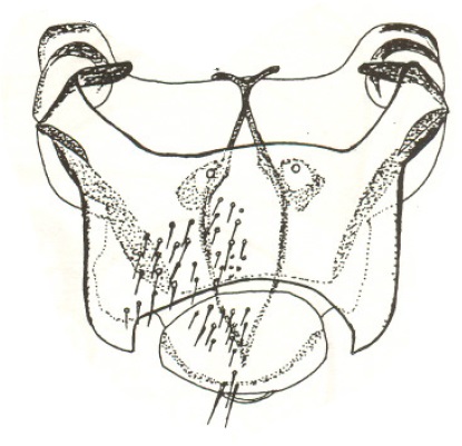 |
| Figure 760. Anapausis soluta, female genitalia in dorsal view |
 |
| Figure 761. Scatopsinae, wings |
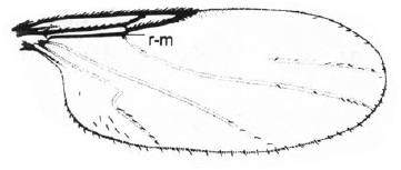 |
| Figure 762. Thripomorpha sp., wing |
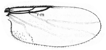 |
| Figure 763. Rhegmoclemina sp., wing |
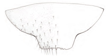 |
| Figure 764. Parascatopse distorta, male sternite 7 |
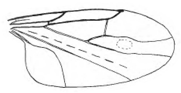 |
| Figure 765. Holoplagia bullata, female wing |
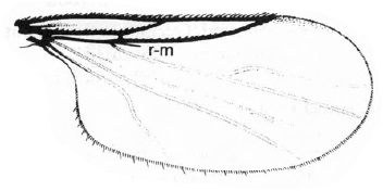 |
| Figure 766. Colobostema variatum, wing |
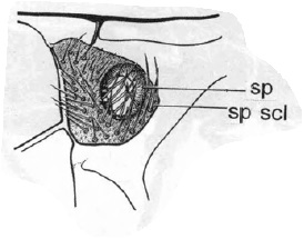 |
| Figure 767. Scatopse notata, anterior spiracle |
 |
| Figure 768. Rhexoza incisa, anterior spiracle |
 |
| Figure 769. Swammerdemella acuta, head in lateral lateral view |
 |
| Figure 770. Pharsoreichertella hamifera, wing |
 |
| Figure 771. Apiloscatopse filamentosa, wing |
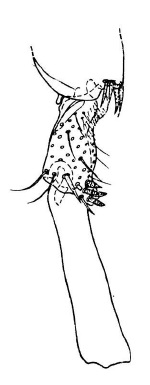 |
| Figure 772. Scatopse globulicauda, basal segments of male hind tarsus |
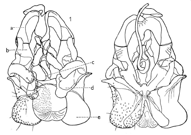 |
| Figure 773. Scatopse globulicauda, male genitalia |
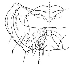 |
| Figure 774. Scatopse globulicauda, female genitalia in dorsal view |
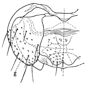 |
| Figure 775. Scatopse globulicauda, female genitalia in ventral view |


